Knock Knee Quick Links: Overview | What is Knock Knee | Is Knock Knee Normal | Causes of Knock Knees | Knock Knee Symptoms | Diagnosing Knock Knees | Treatment
Knock Knee Treatment Videos
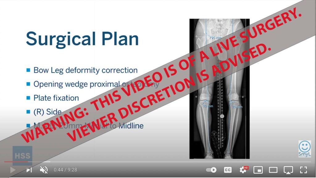
Dr. Rozbruch Discusses Knock-Knee Treatment
Knock Knee Correction Overview
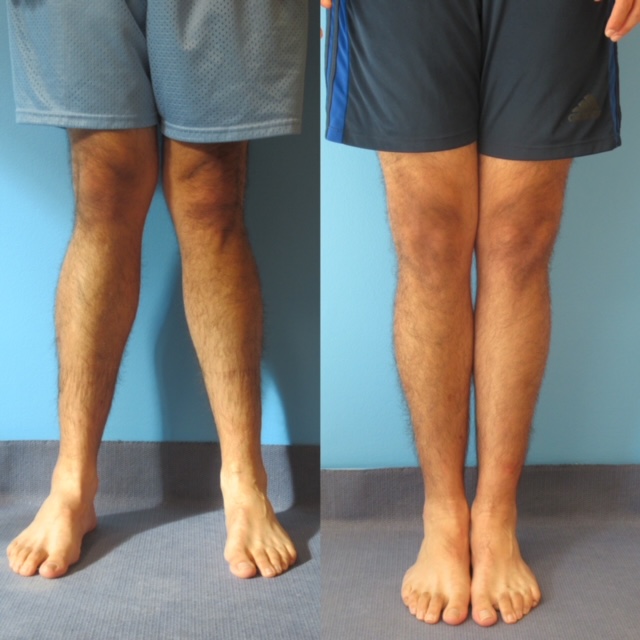
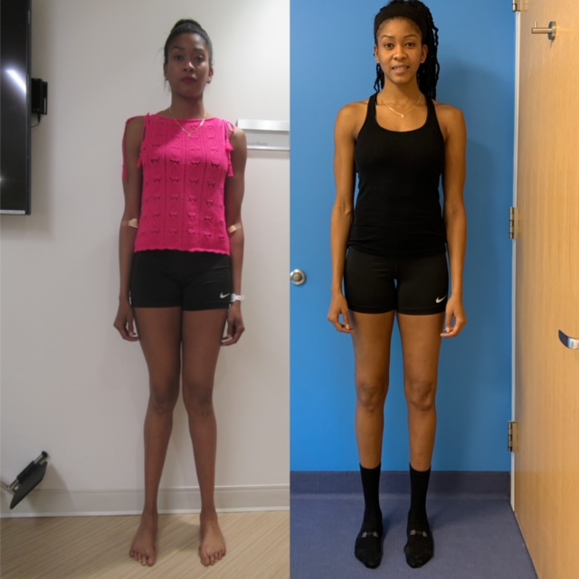
Aiman’s Knock Knees
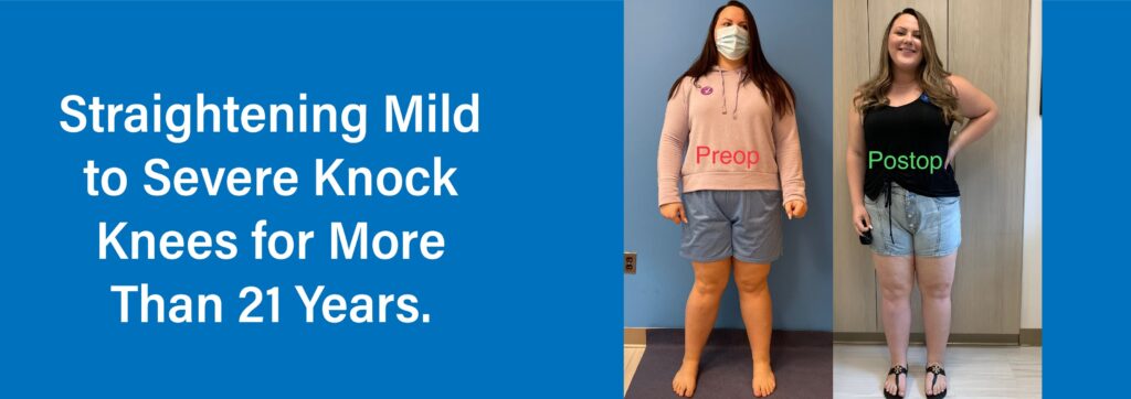
Sandra’s Knock Knee Story
Suzanne was having difficulty with mobility in her socket which was worsened by her valgus #knockknee alignment. I did #knockkneecorrection and Osseointegration Limb Replacement Surgery in one stage. #osseointegrationlimbreplacement https://www.hss.edu/osseointegration-limb-replacement.asp #osseointegration #amputationsurgery #amputee #cyborgsworld @cyborgsworld #amputeelife #amputeestrong #amputation #osseointegrationUSA www.osseointegrationUSA.com #prostheticlimb #prostheticleg #prostheticsandorthotics #limbreplacementsurgery #limbreplacement #osseointegrationaustralia #osseointegrationswholift #Rozbruch #aaos #llrs #orthopedicsurgery #liveyourbestlife @all4ortho #orthopedicsurgeon #hospitalforspecialsurgery #limblengthening @hspecialsurgery @limbdeformity @reifmd #SRobertRozbruchMD www.limblengthening.com www.hss.edu/limblengthening www.hss.edu/LSARC #hssLLCRS #HSSlimbLengthening

Suzanne was having difficulty with mobility in her socket which was worsened by her valgus #knockknee alignment. I did #knockkneecorrection and Osseointegration Limb Replacement Surgery in one stage. #osseointegrationlimbreplacement https://www.hss.edu/osseointegration-limb-replacement.asp #osseointegration #amputationsurgery #amputee #cyborgsworld @cyborgsworld #amputeelife #amputeestrong #amputation #osseointegrationUSA www.osseointegrationUSA.com #prostheticlimb #prostheticleg #prostheticsandorthotics #limbreplacementsurgery #limbreplacement #osseointegrationaustralia #osseointegrationswholift #Rozbruch #aaos #llrs #orthopedicsurgery #liveyourbestlife @all4ortho #orthopedicsurgeon #hospitalforspecialsurgery #limblengthening @hspecialsurgery @limbdeformity @reifmd #SRobertRozbruchMD www.limblengthening.com www.hss.edu/limblengthening www.hss.edu/LSARC #hssLLCRS #HSSlimbLengthening
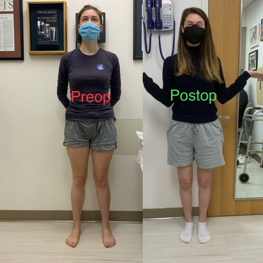
Patient before and after treatment of severe #knockknees #knockkneecorrection and #leglengthdiscrepancy treated with 3 osteotomies using opening and closing wedge strategies to improve #limblengthdiscrepancy #Rozbruch Big improvement in function and confidence and body image #aaos #llrs #orthopedicsurgery #liveyourbestlife @all4ortho #orthopedicsurgeon #hospitalforspecialsurgery #limblengthening @hspecialsurgery @limbdeformity @reifmd #SRobertRozbruchMD www.limblengthening.com www.hss.edu/limblengthening www.hss.edu/LSARC #hssLLCRS #HSSlimbLengthening @usherglanzer #refuahhelpline

Patient before and after treatment of severe #knockknees #knockkneecorrection and #leglengthdiscrepancy treated with 3 osteotomies using opening and closing wedge strategies to improve #limblengthdiscrepancy #Rozbruch Big improvement in function and confidence and body image #aaos #llrs #orthopedicsurgery #liveyourbestlife @all4ortho #orthopedicsurgeon #hospitalforspecialsurgery #limblengthening @hspecialsurgery @limbdeformity @reifmd #SRobertRozbruchMD www.limblengthening.com www.hss.edu/limblengthening www.hss.edu/LSARC #hssLLCRS #HSSlimbLengthening @usherglanzer #refuahhelpline
This young man wasn’t able to get comprehensive medical care as a child so needed a lot of work on his congenital femoral deficiency.
I chose to cut the femur in two places to lengthen, rotate, and correct his alignment. The lengthening #precice nail was able to bend impressively and not break, but the bend changed the alignment. Swift replacement with a solid nail prior to full healing got him straight, just like the plan! Now he is driving to the basket for easy layups!
Finally…we have ChatGPTs interpretation of my cats (see prior posts)
#hssllcrs #hsslimblengthening #limblengthening #knockknee @globusmedicalinc @bodycad_ #femur #deformity #bonedeformity #femurlengthening #orthopaedics #orthopedicsurgery
This is a basic #knockknee correction that so many people would benefit from. Look around and you’ll start to notice this everywhere. If it’s your friend, tell them something can be done, they probably don’t know!
#hssllcrs #hsslimblengthening #knockkneecorrection #orthopedics #orthopedicsurgery @bodycad_ #deformity #deformitysurgery
Guided growth is a technique we can use in children to correct angular deformity without osteotomy. By temporarily stopping growth on one side of joint, the other side can catch up and change the shape of the bones with time (~2 years). The deformity tends to recur, so a slight over-correction is planning to allow for bounce back. This was a large bowleg corrected with minimal surgical exposure!
#hssllcrs #hsslimblengthening #hsskids #bowlegs #knockknees #guidedgrowth #hemiepiphysiodesis #orthopedics #orthopedicsurgery
This is Mr P now, hauling thousand-pound trains of garden supplies eight to ten hours every day. But he couldn`t always do this. When I met him, he had trouble even walking a normal pace in the office. He was born with #fibularhemimelia and had many surgeries to his foot. He then sustained a fracture #limbtrauma shortly before presenting. He was tired of his painful foot and his left #knockknees also was painful. Instead of many more surgeries to his left foot that were uncertain to make him that much better, he opted for #osseointegration. He has felt better with a limb length difference because he has lived his whole life that way. But now with his leg straight and painless, he is the Greek God of Gardening Goods!
.
.
.
#all4ortho #hssLLCRS #HSSlimbLengthening #limbloss #limbdifference #hspecialsurgery
Mr N had left congenital #fibularhemimelia #limbdifference since birth, and then had #limbtrauma and prior surgical care before meeting me. Instead of multiple unpredictable surgeries to save a not-so-good foot, I offered him tibia #amputation with #osseointegration followed by femur osteotomy for #knockknees correction. He has done amazing!!
.
.
.
#all4ortho #hssllcrs #hsslimblengthening #limbloss #hspecialsurgery
Ms A came in with pain in her left leg that was preventing her from working and caring for others. She had not only #knockknees but also an #osteochondroma cartilage growth on her tibia. In a single surgery I removed the osteochondroma and also made her leg straight with a distal femur osteotomy. Here I am showing the intraoperative image of the trajectory for the cut, followed by the bone opening, then getting the alignment straight from the hip to the knee to the ankle, and finally the plate. Here is how her xray looks now, with the left leg perfectly straight! It is amazing how much happier she is now!
.
.
.
.
#all4ortho #hspecialsurgery #hssllcrs #hsslimblengthening
Z and I had a wonderful time fixing her #knockknees
Beyond just straightening her legs and optimizing her knee health, the surgery and follow up allow surgeon and patient to chat about family, careers, delve into what ails the world, and provide reciprocal support and advice. It can make graduation day a bittersweet visit, but with results and memories that last a lifetime!
#hssllcrs #hsslimblengthening #orthopedicsurgery #bonedeformity #bone #knockkneecorrection #femur #osteotomy @bodycad_
This is Ms M. Previously featured on my channel. She is now 6 months out from her right distal femur osteotomy to address severe #knockknees and #kneepain . She struggled with pain that impaired her walking which is much better today! It was amazing to hear how this surgery impacted her life.
Here I`m showing her her own Instagram video!
.
.
.
#all4ortho #hspecialsurgery #hssllcrs #hsslimblengthening
A recent publication from #hssllcrs describing our safety performing #tibia #osteotomy . Techniques and outcomes are summarized. This was published in a @aaos_1 network journal. Coauthors include @limblengthening @limbdeformity @reifmd @adamgeffner
.
.
.
#all4ortho #hspecialsurgery #hsslimblengthening #bowleg #knockknees
This patient of mine presented urgently with a tibia lengthening gone wrong. She had valgus stemming from the femur, lengthening site, and distal tibia! We took things in stages, first salvaging the lengthening, then correcting the femur, and finally the distal tibia. She enjoys seeing her straight leg in photos like this one! #hssllcrs #hsslimblengthening #orthopedicsurgery #tibia #bone #deformitycorrection #knockknees #valgus #bonelengthening #orthopedics #knockknee #bonedeformity

This patient of mine presented urgently with a tibia lengthening gone wrong. She had valgus stemming from the femur, lengthening site, and distal tibia! We took things in stages, first salvaging the lengthening, then correcting the femur, and finally the distal tibia. She enjoys seeing her straight leg in photos like this one! #hssllcrs #hsslimblengthening #orthopedicsurgery #tibia #bone #deformitycorrection #knockknees #valgus #bonelengthening #orthopedics #knockknee #bonedeformity
Overview of the Knock Knee Condition
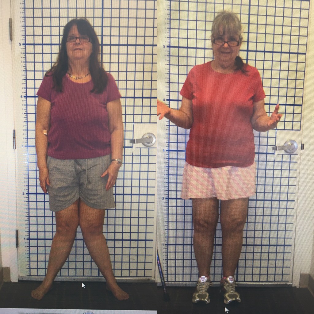
A condition in which the knees bend inward, touching (or “knocking”) even when a person is standing with their ankles apart, knock knee can affect people of all ages. Knock knee, also known as genu valgum, is a mal-alignment of the knee with various causes, often leading to pain and degeneration of the knee if left untreated. Knock-knees can be congenital, developmental, or post-traumatic.
The common theme that pervades all age groups is that the knee is abnormally loaded which can lead to pain, increasing deformity, instability, and progressive degeneration. Correction of the deformity leads to improved knee mechanics, better walking, less pain, and prevents the rapid progression of damage to the knee.
Adult patients who have had knock-knee for many years overload the outside (lateral compartment) and stretch the inside (medial collateral ligament) leading to pain, instability, and arthritis. To prevent and delay the need for joint replacement, knee realignment should be done with osteotomy.
What Is Knock Knee?
Knock knee is a condition in which a person’s knees angle toward each other and touch (“knock”), even when the person is standing with their ankles apart. While temporarily knocked knees is a standard developmental stage in most children, this often self-corrects by age seven or eight. Knock knees that persist beyond six years of age, are severe, or affect one leg significantly more than the other may be a sign of knock knee syndrome.
Knock knee that falls outside the normal developmental patterns may be caused by disease, infection, or other conditions. In these cases, orthopedic intervention may be required to treat the knock knee condition.
Is Knock Knee Normal?
Most children experience normal angular changes in their legs as they grow. When a child is born, they are typically bowlegged until they begin walking at 12-18 months. By the time the child is two to three years old, his legs should begin to angle inward, making him knock-kneed. However, knock-knee usually self-corrects by the time a child is seven to eight years old.
If the angular profile (the leg’s angle from hip to foot) falls outside normal patterns, worsens over time, or is present on only one side of the body, it may be indicative of a more serious form of knock knee requiring further evaluation.
What Causes Knock Knee?
While knock knee is part of a child’s normal growth pattern, persistent forms of knock knee may stem from an underlying disease, infection, or injury. Some common causes of knock knee include:
- Metabolic disease
- Renal failure
- Physical trauma
- Arthritis, particularly in the knee
- Bone infection (osteomyelitis)
- Rickets (a bone-weakening disease caused by lack of vitamin D)
- Congenital
- Growth plate injury
- Benign bone tumors
- Fractures that heal with deformity (malunion)
Being overweight or obese can also put extra pressure on the knees and contribute to knock knee.
Symptoms of Knock Knee Syndrome
The most commonly observed symptom of knock knee is a separation of the ankles when the knees are together. Other symptoms of knock knee often arise as a result of the walking gait adopted by individuals with knock knee. These symptoms may include:
- Pain in knees, feet, hips, and ankles
- A limp when walking
- Stiff or sore joints
- Feet do not touch when standing with knees together
- Knee or hip pain
- Reduced range of motion in hips
- Difficulty walking or running
- Knee instability
- Progressive knee arthritis in adults
- Patients or parents may be unhappy with aesthetics
Other symptoms from underlying conditions may also be present.
How is Knock Knee Diagnosed?
Knock knee is diagnosed through a thorough review of an individual’s medical history, pre-existing conditions, family history, and current health. It also includes a physical examination of the patient’s legs and observations of their gait. Diagnosis also includes a standing alignment X-ray. A standing alignment X-ray or EOS image produces an image of the leg from hip to ankle which helps the orthopedist locate the mechanical axis of the deformity as well as its location. On the x-rays, the magnitude and location of the deformity is identified.
How Is Knock Knee Treated?
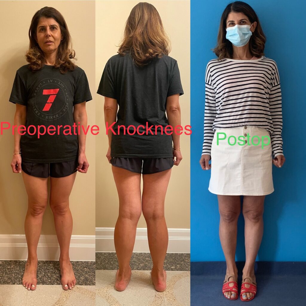
Treatment for mild cases of knock knee in children or adolescents may include braces to help bones grow in the correct position. If a gradual correction does not occur, surgery may be recommended.
In the growing child, guided growth minimal incision surgery may be used to encourage the limb to gradually grow straight. Osteotomy, in which the femur is cut, and then realigned may be needed. In some cases, the surgeon also places an external fixator. With external fixators, pins are inserted into the bone and protrude out of the body to attach to an external stabilizing structure. Physical therapy is also an important part of treatment, especially in cases where surgery has occurred.
In skeletally mature adolescents and in adults, osteotomy is the recommended treatment to straighten the leg. X-rays are used to determine the location and magnitude of the deformity. In most cases, we treat the femur, but there are situations when the tibia or both femur and tibia are treated. When moderate deformity is present, the osteotomy is typically stabilized with internal fixation (plate or rod). When the mal-alignment is more severe, gradual realignment of the limb through the osteotomy may be done with an external fixator.

